Pets commonly injure their cranial cruciate ligament, and typically require surgical intervention to eliminate the joint instability. Our team at Premier Pet Hospital wants to help you understand tibial plateau leveling osteotomy (TPLO), so you will know what to expect if your pet is affected.
Cranial cruciate ligament rupture in pets
The femur and tibia are held together by cruciate ligaments to form the stifle (i.e., knee joint). The cranial and caudal cruciate ligaments cross each other in the middle of the joint to stabilize the stifle, creating a hinge joint. Because this construction does not include any interlocking bones, the stifle is relatively unstable, and commonly injured. The cranial cruciate ligament is more commonly injured in two ways:
- Twisting injury to the knee — When a running pet suddenly changes direction, they place the majority of their body weight on the knee, causing excessive rotational forces on the cruciate ligaments. When the ligament—most commonly the cranial cruciate ligament—ruptures, the joint becomes unstable, causing the pet significant pain and lameness.
- Cranial cruciate ligament disease (CrCLD) — CrCLD is the most common cause of cranial cruciate rupture in dogs, and is caused by several factors, including ligament degeneration, obesity, poor physical condition, genetics, conformation, and breed. CrCLD is a subtle, slow degeneration, rather than a sudden traumatic event. Certain breeds, such as rottweilers, Labrador retrievers, Chesapeake Bay retrievers, and mastiffs, are at increased risk for CrCLD. About 40% to 60% of dogs who have CrCLD in one knee will develop the condition in the other knee, and a dog may experience a partial cranial cruciate ligament tear that eventually progresses to a full tear.
History of the tibial plateau leveling osteotomy in pets
In the early 1960s, veterinarians attempted to replace the damaged ligament after a cranial cruciate ligament rupture, but the procedures did not completely stabilize the injured joint, and led to severe arthritis. In the 1980s, Dr. Barclay Slocum examined the knee joint’s structure and its dynamic forces during weight bearing and movement. He noted that the tibia slopes from front to back inside the knee, with a 21- to 24-degree tibial plateau slope in most dog breeds. He also noted that when the cranial cruciate ligament was ruptured, the femur slid down the tibial slope, and he proposed that pets were most affected by this instability. He theorized that leveling the slope would more successfully stabilize the joint, and devised the surgical technique called the TPLO.
The tibial plateau leveling osteotomy procedure in pets
To perform a TPLO, an X-ray is taken of your pet’s stifle joint, and the angle at the top of the shin bone (i.e., the tibial plateau angle) is measured. The procedure’s goal is to reduce this angle so the tibia can’t slide forward, which creates joint instability. The stifle joint first is assessed to ensure healthy meniscus cartilage, because any damaged cartilage must be removed before the TPLO can proceed. The TPLO procedure involves making a curved cut on the inside surface of the top of the tibia with a curved saw blade, rotating the cut portion to create the desired tibial plateau angle, and placing a stainless steel plate to hold the pieces in their new alignment.
Pets who benefit from tibial plateau leveling osteotomy
Only about 15% of pets will recover acceptable function without surgery, and these pets typically weigh less than 15 to 20 pounds. A TPLO can be performed on any pet, including small dogs and cats, and is the best choice for dogs who weigh more than 45 pounds, especially if they are active. If the joint isn’t properly stabilized, arthritis will develop, causing pain and decreased mobility for your pet.
Post-operative care for tibial plateau osteotomy in pets
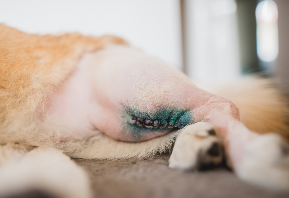
Your pet’s activity will be restricted after surgery because premature, uncontrolled, or excessive exercise can cause complete or partial surgical repair failure. Physical rehabilitation can help speed your pet’s recovery and improve their final outcomes, and our veterinary professionals will customize a plan for your pet. TPLO healing is typically rapid:
- 24 hours post-surgery — About 50% of patients will begin bearing weight on the injured limb.
- Two weeks — Most pets can bear moderate to complete weight on the limb.
- 10 weeks — Most pets have no noticeable limp or gait change.
- Four months — Most pets can begin walking and playing normally.
- Six months postoperatively — The majority of pets can resume full physical activity.
Preventing cranial cruciate ligament rupture in pets

Not all cranial cruciate ligament ruptures in pets can be avoided, but you can lower your pet’s risk. Steps include:
- Keeping your pet at a healthy weight — Overweight pets are at higher risk for cranial cruciate ligament rupture, partly because the excess weight puts more pressure on their joints. If your pet needs to lose a few pounds, ask our veterinary professionals to help you devise a safe weight loss strategy.
- Providing regular exercise — Ensure your pet gets daily exercise to keep their joints and ligaments strong and healthy, and to help them maintain a healthy weight.
- Monitoring your pet for gait abnormalities — If your pet’s cranial cruciate ligament starts to deteriorate, the condition should be treated before the ligament fully ruptures, to prevent further damage.
Cranial cruciate ligament rupture is a painful condition that can greatly decrease your pet’s mobility, but our veterinary professionals have the skill and training to perform a tibial plateau leveling osteotomy to resolve the problem. If your pet is limping, contact our team at Premier Pet Hospital, so we can discover what is causing their lameness and alleviate their pain.


Leave A Comment